Research Article | | Peer-Reviewed

The Effects of Electroconvulsive Shock on the Superior Colliculus Visual Evoked Potential

Received: 24 April 2025     Accepted: 8 May 2025     Published: 16 June 2025
Views:       Downloads:
Abstract

Electroconvulsive therapy (ECT) remains an effective somatic treatment for a variety of psychiatric disorders. Despite its introduction almost a century ago, its site and mode of action remains elusive. One method of obtaining relevant information is by the recording of sensory evoked potentials from patients undergoing ECT. However, such human data is plagued with problems of methodology and interpretation. These limitations are not an impediment when employing an animal model of electroconvulsive shock (ECS). In the present experiment, the effects of ECS were studied on the superior colliculus visual evoked potential (SCVEP) in the non-medicated rat. The SCVEP was used as a measure of activity in the subcortical visual system as, in the rodent, the SC lies directly below the visual cortex. Immediately after the induction of generalised seizure activity (GSA) by ECS, all the components of the SCVEP were still preserved, basically intact. There was, however, a marked but very transient attenuation in its waveform not associated with any increase in the latency of the primary component. These findings are compared to a previous study where the cortical VEP was found to be completely abolished for up to two minutes following ECS. Judging solely by these and related neurophysiological data recorded from other exteroceptive sensory systems, it is concluded that the seat of ECT’s mode of action may lie quite discretely at the cortical level. Nevertheless, ECT has a profusion of effects on cerebral structure and function. Therefore, at the present time, this putative insight may be most applicable to an understanding of the negative or adverse (usually cognitive) side-effects which accompany ECT rather than to its positive (therapeutic) benefits. The acute loss of amplitude in the SCVEP waveform may not necessarily indicate that GSA impacts SC function, even momentarily. Instead, it is suggested that the interference more likely reflects a deficit in retinal processing which has been transferred to the SC.

Published in American Journal of Psychiatry and Neuroscience (Volume 13, Issue 2)
DOI 10.11648/j.ajpn.20251302.13
Page(s) 61-71
Creative Commons

This is an Open Access article, distributed under the terms of the Creative Commons Attribution 4.0 International License (http://creativecommons.org/licenses/by/4.0/), which permits unrestricted use, distribution and reproduction in any medium or format, provided the original work is properly cited.

Copyright

Copyright © The Author(s), 2025. Published by Science Publishing Group

Keywords

Electroconvulsive Shock, Electroconvulsive Therapy, Flash Visual Evoked Potential, Gap Junction, Generalized Seizure Activity, Superior Colliculus, Occipital Cortex

1. Introduction
Electroconvulsive therapy (ECT), where an electric current is used to induce an artificial state of generalized seizure activity (GSA), remains an effective, if still controversial, treatment for a range of psychiatric illnesses. Most notably, these include endogenous depression, manic – depression, mania, catatonia and schizophrenia . It may also alleviate the symptoms of movement disorders such as Parkinson’s disease . ECT has a number of monikers. In psychiatry, it is also known as electroshock treatment, shock treatment, electroplexy and electrocerebral therapy . In veterinary practice it is called electrical stunning. In animal research it is normally labelled electroconvulsive shock (ECS).
Despite its longstanding use, the site and mode of action of ECT is still speculative and it is therefore classified as an essentially empirical treatment. Nonetheless, since its introduction in the 1930s, well over 100 theories have been proposed to explain its beneficial clinical effects although none has proved totally satisfactory . Such explanations can be roughly categorized as neuropsychological, neurophysiological, neuroendocrinological, neurochemical, neuroplastic, neuroinflammatory and neuroimmunolgical models . The bulk of the neurophysiological research has involved quantifying the effects of ECT on the electroencephalogram (EEG) during a course of treatment . However, during the 1970s and 1980s, this information was complemented by several attempts to record sensory evoked potentials (EPs) from the visual, auditory and somatosensory systems following ECT in psychiatric patients . While providing some useful information, this data was compromised by methodological limitations and confounding effects of concurrent medication. So far as can be determined, no further studies have been reported for almost four decades.
Most of the shortcomings of these clinical recordings can be overcome with an animal model of ECS where a wider range of exteroceptive potentials can be recorded than with human subjects . Summarizing these findings, it was discovered that while cortical waveforms were invariably but transiently lost immediately after ECS, potentials generated elsewhere in the brain were always preserved basically intact. This included subcortical potentials recorded from both the auditory and somatosensory systems. However, what was still missing from this data set was a measure of the impact of ECS on subcortical visual activity.
In the present experiment, therefore, the effects of ECS were examined on the superior colliculus visual EP (SCVEP) in the rat. In the rodent, the SC is a comparatively large structure which lies directly beneath the occipital lobe and retinal images are projected in a topographical manner onto both . The SC is innervated by the brachium of the SC which is a collateral pathway branching off the optic tract just prior to the waystation of the lateral geniculate body . In the rodent, a large proportion of optic pathway fibers (possibly 50% or more) have the SC as their destination and therefore switch direction at this point . This is an indication of the pivotal role that the SC continues to play in guiding visuo-motor activity in rodents.
The SCVEP can be readily accessed using a needle electrode inserted through a trephine hole and this is probably the reason for its popularity when studying the effects of various agents and conditions on SCVEP function in the rat. Examples include the effects of air pollution , insecticides , carbon monoxide , pentobarbital , alcohol , ketamine , naloxone and physostigmine , GABA agonists , anticonvulsants , nicotine , muscle relaxants , amphetamines , antihypertensives and CNS stimulants . However, the present account is the first description of the effects of GSA on the SCVEP.
2. Materials and Methods
Subjects were 10 adult male albino rats (300-350 g). Approximately one week prior to the experiment, each animal was anesthetised with pentobarbital (60 mg/kg). The skull was exposed and a trephine hole was drilled directly over the left SC (1.5 mm lateral to the sagittal suture and 6 mm posterior to bregma . A small rubber cap was placed over the hole and anchored in position with dental acrylic. Two skull screws were implanted sequentially along the nasal bone and also secured with dental cement.
On the experimental day, each animal was initially curarized with a dose of d-tubocurarine chloride (4 mg/kg). As soon as signs of neuromuscular paralysis became apparent, the subject was connected to a respirator via a rubber mask anchored to the more distal nasal bone screw and artificially ventilated at a rate of 50 strokes per minute so as to maintain a normal heartrate (350-400 BPM). This technique was an adaptation of that originally used by Miller and co-workers during their classical studies of biofeedback . The indifferent lead was connected to the more proximal of the nasal bone screws. The ground lead was connected to a needle electrode inserted through the nape of the neck. The active electrode was a stainless steel needle insulated except for a bared 1 -2 mm tip. It was inserted through the rubber cap and lowered approximately 4 mm from the top of the skull so that its tip penetrated the superficial layers of the SC .
SCVEPs were recorded using a Tracor-3000 evoked response analyser. Analysis time was 100 msec and the sampling interval 200 µsec. Bandpass of the amplifiers was set to 1-250 Hz. Thirty two responses were averaged to obtain each SCVEP. The stimuli were light pulses delivered by a Grass model PS 22 photic stimulator. Duration of each flash was 10 µsec and stimulation rate was 2/sec. Flash intensity was set to step 8 on the 1-16 scale. Recordings were made following monocular stimulation of the right (contralateral) eye in a darkened room.
Initially, the optimum SCVEP was obtained by carefully adjusting the depth of the active electrode. The SCVEP has a very distinctive waveform consisting of five main subcomponents (Figure 1) and has a quite different morphology from the cortical VEP generated directly above it. Recording a reproducible and characteristic SCVEP was used as the baseline and also as confirmation that the needle electrode was located in the SC.
GSA was induced by transmitting a brief electric current (80 mA for 600 msec) via miniature bulldog clips attached to the ears. The inside of the clips were smeared with electrode paste. This magnitude of ECS will induce tonic-clonic seizures in the awake non-paralysed animal lasting approximately 1 minute associated with a period of insensitivity and loss of reflex activity for up to 3 minutes.
The first SCVEP was made between 0 – 30 seconds (0 minutes). A second recording was made between 30 – 60 seconds (1 minute) and subsequent SCVEPs were obtained at 1 minute intervals until 10 minutes. The animal was then immediately euthanized with an overdose of pentobarbital.
The use of neuromuscular paralysis by curariform agents has two possible functions. First, as a kind of faux anesthetic on the false assumption that because an animal is immobilized it must also be insensible. Such a procedure has long been considered unethical and therefore proscribed. The second potential use is simply as a chemical restraint and therefore should not be conflated with the first. This technique has been widely used in both human e.g. and animal e.g. experimentation and there is no reason or evidence to believe that it is in any respect a painful or stressful experience. It is also notable that β -endorphin plasma concentrations do not increase during neuromuscular blockade .
The project was approved by the University of Auckland Animal Ethical Committee (AEC no. 324).
3. Results
Two examples of the normal SCVEP recorded from the awake rat are shown in the baseline (Before ECS) example in Figure 1. Initially, there is a complex consisting of two positive subcomponents usually labelled P1 and P3. This is followed by a negative trough (N4) which separates a secondary positivity (P4) followed by a late negative response (N5). These five subcomponents are identified in the baseline example of subject 1 (Figure 1). This basic waveform has been frequently described in the rat . In the present study, only the activity of the primary collicular component (P1) was formally analysed. It is understood that P1 reflects post-synaptic activity generated following the arrival of fast-conducted impulses of the optic stratum .
Two examples of the effects of ECS on the SCVEP are illustrated in Figure 1. It can be seen that, apart from a general diminution of voltage, ECS otherwise had little or no impact on the waveform. Even during the acute ictal phase (0 min), all the subcomponents of the SCVEP were clearly present. Despite the attenuation in amplitude, there was no concomitant increase in the latency of P1, as might otherwise have been expected. The loss of amplitude was also quite transitory and was not obviously apparent beyond the fourth post-ECS recording at 3 minutes.
Mean latency and amplitude data for the P1 component is summarised in Figure 2. This confirms the individual findings in Figure 1 that P1 was always preserved after ECS albeit with a substantial loss of amplitude but no evidence of an overall increase in latency. Mean voltage of P1 sank to less than one half of its baseline value immediately after ECS but then rapidly recovered within the next 5 minutes. However, the amplitude subsequently remained static and was never fully restored during the remainder of the recording period. This is in contrast to the two examples illustrated in Figure 1.
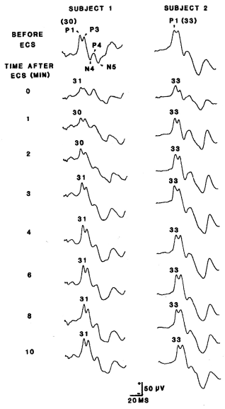
Figure 1. Two examples of the effects of ECS on the SCVEP. In the baseline examples, the primary collicullar component (P1) is identified with its actual latency (ms) in parentheses. In the post – ECS SCVEPs, only the latency of P1 is indicated. SCVEPs recorded at 5, 7, and 9 minutes are not illustrated. In the baseline recording of subject 1, all 5 principal components of the waveform are identified.
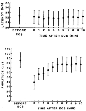
Figure 2. Mean latency and amplitude (± 1 SD) of the P1 component of the SCVEP at the times indicated following ECS. The amplitude of P1 was calculated by referring it to the shallow negative trough preceding P1.
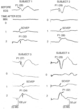
Figure 3. Four examples of the effects of ECS on the cortical VEP. The primary cortical response (P1) is indicated with the arrows and the actual latency is in parentheses. Note the absence of the waveform at 0 minutes but the appearance of a low amplitude positivity at 1 minute. This is a far field recording of the SCVEP, not the cortical potential. Note also the enhanced amplitude of P1 in the post-ictal recording at 2 minutes. Details on how these EPs were recorded are available elsewhere . Illustrations of the waveforms were made using a photochemical process and in the interim the quality of them deteriorated and faded making reproduction less than optimal. In some traces (especially subject 1), either the beginning or end is missing but the latency values are accurate. The scale at the bottom left applies to both subjects 1 and 3 while the bottom right scale applies to subjects 2 and 4.
4. Discussion
In lower mammals, such as rodents, the SC operates as a primitive visual receiving area rather than just an optic reflex center . As presently demonstrated, it also generates a robust high amplitude VEP which is not masked or otherwise interfered with by far field retinal activity . As discussed in the Introduction, the SCVEP is not directly generated as part of a sequence within the retino-geniculo-cortical pathway. The anatomical organisation of the retino-collicular tract means therefore that the SCVEP is only a partial measure of the activity propagated within the retino-geniculo-cortical pathway. Findings with respect to the effects of ECS on the SCVEP may consequently need to be interpreted with a degree of caution.
Nevertheless, the evidence that a subcortical visual waveform can persist immediately after the induction of GSA by ECS is consistent with the findings in the somatosensory and auditory systems . They are also in some respects quite remarkable. This is because, unlike the thalamus, the SC in the rodent is the closest of neighbors to the visual cortex. Despite this, activity in one structure (the occipital cortex) is totally abolished or suppressed during a state of GSA while in the other (the SC) it is maintained during the ictal period with only a very transient alteration in its waveform. This is another example in neuroscience where the significance of negative findings can often be misunderstood or underrated.
This quite distinctive discrepancy can be more clearly appreciated by comparing Figure 2 with Figure 3. In Figure 3, four examples of the effects of ECS on the cortical VEP are displayed. In contrast to the SCVEP, these illustrations demonstrate how the cortical waveform is totally destroyed by ECS/GSA (0 minute). However, at 1 minute, a small positive potential or notch can almost always be discerned. This is not the reappearance of the cortical potential. Rather, it is the far field recording of the SCVEP . The SCVEP is normally masked by the cortical VEP but becomes temporarily visible during the early post-ictal interval when the cortical waveform is still absent. Note two characteristics of the cortical VEP upon its initial return at about 2 minutes. First, the latency of the primary cortical response (P1) is prolonged thereby indicating that the diminutive waveform recorded at 1 minute cannot be of cortical origin. Second, the P1 component is also abnormally enhanced.
The present results provide the final part of a neurophysiological jigsaw. Each part represents the effects of ECS on a particular exteroceptive EP recorded from the surface of the rat brain. Many of these waveforms can also be recorded from the scalp of humans although with some notable exceptions including the SCVEP. The SCVEP is an outlier for another reason. This is because it is located directly below the visual cortex which normally acts as a barrier for recording from a surface electrode. This means that an unorthodox technique needed to be employed to access it.
The overall picture that emerges of how the brain responds to ECS/GSA is quite simple and straightforward. Cortical activity in all three exteroceptive sensory systems is instantly and completely knocked out albeit for a brief period of time but normal function is rapidly restored. In marked contrast, activity generated in subcortical, thalamic, collicular, cerebellar and brainstem tracts and nuclei survives even during the acute ictal phase. There are only minor alterations to the waveforms although once again the SCVEP was an outlier in this regard. This anomaly will be subsequently considered.
According to standard doctrine, EPs recorded from scalp or surface arise in one of two possible generators . One source is the compound action potential (CAP) exemplified by the high frequency sharp waves of the brainstem auditory evoked potential (BAEP). More commonly, however, the EPs reflect graded post-synaptic activity originating predominantly in dendrosomatic locations. These can be distinguished by their slow frequency content which is rarely greater than 300 Hz. The SCVEP is one such example. There is also unanimity that postsynaptic potentials (PSPs) (both excitatory and inhibitory) are the exclusive origin of EPs generated in the cortex.
The present analysis of the effects of ECS on EPs provides additional insights into both normal and abnormal neuronal function. First, the wave of depolarisation which sweeps down an axon thereby generating a CAP is in no way hindered or impeded by a state of GSA. Nor does it prevent the diffusion of chemical neurotransmitters across the synapse. Otherwise, the EPs dependent upon these processes could not still have been generated usually unimpaired. Conversely, the immediate abolition of the cortical waveforms is quite contrary to the common understanding that they are generated by PSPs. These, as discussed above, seem generally resistant to degradation by ECS/GSA. It follows, therefore, that cortical waveforms must have a quite distinct electrogenesis from that which is conventionally assumed. If this is the case, the only alternative candidate must be that the cortical EPs are being generated by electrotonic processing via electrical synapses.
With electrical coupling, cell communication with neighbours is conducted via gap junctions . Gap junctions consist of a pair of hemi-ducts or connexons. Each connexon is composed of a cluster of six cylindrical protein molecules or connexins. One of the pair protrudes from the presynaptic membrane and the other from the postsynaptic membrane. When joined together, they form a central channel or pore. This arrangement allows positively charged sodium ions (Na+) to flow efficiently and nearly simultaneously through the pipeline. This activity evokes an excitatory PSP in the postsynaptic cell thereby potentially depolarizing the neuron. It stands to reason that a train of ions traversing a gap junction would be more susceptible to a paroxysmal burst of electrical energy than would a diffusion of a neurochemical released into extracellular fluid.
However, the afferent signals crossing the gap are essentially just an extension of the CAP. As discussed above, this activity also seems immune to GSA. A more feasible mode of action might therefore be the instantaneous closing of the gap junction gate under the influence of the abnormal electrical seizure activity. This would result in the sudden and total blockage of the stream of ions through the hexagonal tunnel and therefore the abrupt loss of the cortical potential in the postsynaptic neuron.
In summary, during GSA, afferent impulses can traverse the central pathways and synaptic relays with impunity or with little or no impedance. However, they are subsequently extinguished or obstructed at the cortical level. This principle would therefore seem to imply that the site and mode of action of ECT might reside at the cortical level and involve disruption of electrical synapses. In this respect, it may be instructive to compare clinical recordings using psychiatric patients with the animal data.
Initially, Small and co-workers recorded longer latency cortical VEPs and auditory EPs (AEPs) during the interictal period, typically 1 – 2 days after a trial of ECT. Only minor and ephemeral changes were reported to the waveforms such as increases in latency and decreases in amplitude of some components. Little information was imparted on the nature of the EPs nor any illustrations provided. In a follow-up study, Small recorded the same AEPs and VEPs from patients much sooner after ECT, typically 1 -3 hours. Again, the authors could detect no significant change to the waveforms apart from an increase in amplitude of the AEPs and no illustrations were provided. The findings of Small and co-workers were largely replicated by Kolbeinsson and Petursson . They recorded long latency AEPs following click stimulation 60 – 90 minutes after ECT but did not observe any alterations or abnormality in waveform.
Somewhat more edifying and instructive is the research of Kriss and co-workers who managed to record shorter latency cortical EPs during the acute ictal period immediately after the induction of GSA by ECT. In their first study , early components of the cortical somatosensory EP (SEP) were recorded beginning a few seconds after the delivery of ECT. Even during the ictal period, the primary cortical component of the waveform could still be identified although the later components were abolished. These were quickly restored although some abnormalities lingered for at least 30 minutes.
In a complementary study , the authors recorded the cortical VEP under the same conditions as the SEP. As in the first experiment, unilateral ECT was employed and this produced complex differences between the two studies. Generally, however, ECT seemed to have more profound effects on the VEP waveform which was essentially suppressed and perhaps lost during the ictal period although there was some evidence that the primary cortical response could still be intermittently registered. Between 3 – 8 minutes, all the components had returned and by 30 minutes the waveform was normal.
There have also been two studies of the effects of ECT on the BAEP which were reported almost simultaneously. Weiner and co-workers recorded BAEPs from 0 – 60 minutes after ECT. Even during the immediate ictal period, no alteration in waveform was found. In the contemporary but less detailed project, BAEPs were recorded at times before, during and after a course of ECT . Little or no change in waveform morphology could be detected during this period. The authors concluded that the BAEP was generally invulnerable to the chronic effects of ECT.
Reviewing most of this data on human EPs and ECT, Weiner concluded that they were “indicative of a transient state of global cerebral dysfunction”. This is not quite a fair assessment. In fact, the findings do suggest that cortical activity is more vulnerable to ECT than that generated in more caudal parts of the brain. This conclusion is therefore more or less compatible with the more detailed animal studies reviewed previously. Presumably, the apparent survival of some cortical components during the ictal phase was likely due to the anti-convulsant properties of the concurrent barbiturate anesthesia raising the seizure threshold.
ECT has multiple effects on different locations and activities in the brain and determining which are responsible for its beneficial qualities has proven difficult to sort out . Further, it is still unclear whether there is a solitary or exclusive therapeutic mechanism operating or whether individual neuropsychiatric disorders must be accounted for by quite separate and different modes of action. Among the many suggestions on how ECT may work are interference with protein synthesis, changes in cerebral metabolism and alterations in cerebral blood flow. Increase in the permeability or rupture of the blood brain barrier is another common idea thereby allowing the influx of a chemical agent with potential anti-depressant and other properties. Somewhat related is the concept that inhibitory anti-convulsant activity designed to quell the GSA may also possess therapeutic efficacy. The synthesis of almost all neurotransmitters is affected by ECT, most particularly biogenic amines such as norepinephrine, serotonin and dopamine. There is widespread release of neuropeptides, neurohormones and neuromodulators, particularly involving the hypothalamus. There are also processes of neuroplasticity involving neurogenesis, dendrogenesis, synaptogenesis, gliogenesis, neuronal sprouting and receptor sensitivity and density. Responsiveness in the immune system and epigenetic mechanisms may also play a role. Any direct or suspected relevance of this heterogeneous collection of putative modes of action of ECT to the present analysis of EPs must at this stage remain conjectural.
There is, however, one possible exception to this conclusion. This is the diencephalic model of ECT action. This theory argues that it is the stimulation of diencephalic structures, either directly by electric current or via GSA which is the sine qua non for the alleviation of affective disorders . Among the evidence adduced is that the passage of bilateral ECT passes straight through the diencephalon unlike unilateral ECT which it is therefore claimed is a less potent treatment. It has also been proposed that the thalamus houses a subcortical pacemaker responsible for controlling cortical rhythmic activity . The progressive slowing of the EEG during a course of ECT is therefore consistent with the concept that ECT may specifically target and therefore activate the diencephalon. The diencephalic model is complemented by the neuroendocrine theory . This contends that diencephalic stimulation and excitation precipitates the release of hypothalamic neuropeptides and other hormones ultimately leading to the relief of the vegetative and homeostatic symptoms of endogenous depression.
It might be predicted therefore that if the assumptions of the diencephalic theory are correct, then ECT could also target EPs arising from the thalamus. Nevertheless, judging by thalamic EPs, there is no evidence that ECT interferes with or influences diencephalic activity . This is not, of course, a definitive inference as rectification of abnormal or unbalanced hypothalamic function could still be mediated via local neuronal circuits whose activity is not detectable using relatively gross EP methodology. Despite the EP findings, the neuroendocrine – diencephalic theory continues to be an important model of ECT action .
In summary, at the present time it is uncertain what the findings of ECT/ECS on EPs might contribute to an overall understanding of its positive or therapeutic properties. Conversely, it seems a reasonable possibility that this data may well help explain the adverse negative side-effects generated by the treatment. These characteristically concern the cognitive defects particularly the short term memory disturbances which typically accompany a course of ECT and are sometimes designated a category of brain damage . Be that as it may, the interference with the consolidation process following ECT continues to be difficult to explain .
In any case. It has been argued that the primary motivation for conducting such studies is not principally to gain insights into the site and mode of action of ECT. Rather, it is because it provides an experimental model with which to study the effects of ECS/ECT on neurophysiological activity . It is clear that this paradigm, especially when employing an animal model, has proven capable of revealing novel information concerning what Weiner called the “acute electrophysiological correlates of generalized seizure activity”.
What can also be stated with some assurance are two principles regarding the interpretation of EPs in clinical practice. The first corollary is that if a cortical waveform recorded during the acute post-ictal period is abnormal, then this is most likely due to the state of GSA. The second corollary is that if a brainstem, midbrain or subcortical EP recorded during the post-ictal period is abnormal, then it is most likely due to some other disorder of cerebral function rather than GSA. This is an elaboration and extension of the rule originally formulated by Weiner .
A final minor but puzzling problem with the present findings concerns the atypical behavior of the SCVEP waveform after ECS. There is no ready explanation for the very brief loss of voltage in its waveform. The SCVEP might normally be augmented by activity transmitted via cortico – tectal pathways. If the SC is temporarily deprived of such innervation following the loss of the cortical waveform, then a significant reduction in amplitude might ensue. However, experiments where the visual cortex was ablated provide little support for this notion. On the face of it, it also seems unlikely to reflect some kind of retinal deficit because it has been reported that the electroretinogram (ERG) was preserved mostly unaltered during GSA . In fact, retinal contribution to the loss of amplitude cannot be ruled out. This is because it is well established that electrotonic activity does play a key role in the processing and integration of visual information within the eye and gap junctions operate at all levels within the retina . It is therefore feasible that a reduction in retinal energy could have been transferred to and therefore reflected in the SCVEP waveform. However, any such impairment might have been masked and therefore gone undetected because of the technical difficulties encountered when attempting to record an optimum ERG during GSA . It may also account for the anomalous dissociation between amplitude (decrease) and latency (no change) of the waveform immediately after GSA. It is almost invariably the case that a reduction in amplitude is correlated with an increase in latency of the SCVEP waveform e.g. . This also implies that it may not have been just at the cortical level that EPs are susceptible to the effects of ECS/ECT. Finally, it is apparent from Figure 3 that the cortical VEP waveform is lost for up to 2 minutes following ECS. This is twice as long as for the cortical SEP and cortical AEP . This is therefore further evidence that there may be more than one site of impact of ECS/GSA within the visual system.
5. Conclusions
1. The recording of EPs following ECT/ECS may be a useful technique for studying the electrophysiological correlates of GSA.
2. According to such findings, an afferent signal can be conducted within the central sensory pathways uninterrupted until it is irrevocably blocked at the cortical level.
3. It can therefore be inferred that cortical EPs may be generated by electrical rather than chemical synapses hence their susceptibility to ECS/GSA.
4. Considering just the neurophysiological recordings from both experimental animals and psychiatric patients, it can be deduced that the site and mechanism of action of ECT may lie within the cortex.
5. ECT has multifarious effects of CNS structure and function. Therefore, at the present stage of knowledge, the preceding insight may be more usefully restricted to an understanding of the cognitive impairment which commonly accompanies ECT rather than of its therapeutic properties.
6. The findings on the effects of ECT/ECS on EPs also allow a rule to be formulated regarding the interpretation of clinical EPs during an ictal state. In summary, this states that if an abnormal subcortical, midbrain or brainstem waveform is recorded under such conditions, it is most likely due to a disorder of cerebral function. Conversely, if an abnormal cortical waveform is recorded, it is most likely due to the GSA.
Abbreviations

AEP

Auditory Evoked Potential

BAEP

Brainstem Auditory Evoked Potential

CAP

Compound Action Potential

ECS

Electroconvulsive Shock

ECT

Electroconvulsive Therapy

EEG

Electroencephalogram

EP

Evoked Potential

ERG

Electroretinogram

GSA

Generalized Seizure Activity

PSP

Post Synaptic Potential

SC

Superior Colliculus

SCVEP

Superior Colliculus Visual Evoked Potential

SEP

Somatosensory Evoked Potential

VEP

Visual Evoked Potential

Author Contributions
Nigel Alexander Shaw is the sole author. The author read and approved the final manuscript.
Funding
This research was supported by the Maurice and Phyllis Paykel Trust.
Conflicts of Interest
The author declares no conflicts of interest.
References
[1] Kalinowsky, L. B., Hippius, H. Pharmacological, convulsive and other somatic treatments in psychiatry. New York: Grune and Stratton; 1969.
[2] Sainsbury, M. J. Key to psychiatry. Sydney: ANZ Book Company, 1973.
[3] Crowe, R. R. Electroconvulsive therapy- A current perspective. The New England Journal of Medicine. 1984, 311, 163-167.
[4] Weiner, R. D. Does electroconvulsive therapy cause brain damage? The Behavioral and Brain Sciences. 1984, 7, 1-53.
[5] Kaplan, H. I., Sadock, B. J., Grebb, J. A. Kaplan and Sadock’s synopsis of psychiatry: Behavioral sciences/ clinical psychiatry. 7th edition. Baltimore: Williams and Wilkins; 1994.
[6] Bolwig, T. G. How does electroconvulsive therapy work? Theories on its mechanism. Canadian Journal of Psychiatry. 2011, 56. 13-18.
[7] McCall, W. V., Andrade C., Sienaert, P. Searching for the mechanism(s) of ECT’s therapeutic effect. The Journal of ECT. 2014, 30, 87-89.
[8] Sienaert, P. Mechanisms of ECT: Reviewing the science and dismissing the myths. The Journal of ECT. 2014, 30, 85-86.
[9] Sackeim, H. A. Modern electroconvulsive therapy: vastly improved yet greatly underused. JAMA Psychiatry. 2017, 74, 779-780.
[10] Singh, A., Kar, S. K. How electroconvulsive therapy works? understanding the neurobiological mechanisms. Clinical Psychopharmacology and Neuroscience. 2017, 15, 210.
[11] Kirov, G., Jauhar, S., Sienaert, P., Kellner, C, H., Mc Loughlin, D. M. Electroconvulsive therapy for depression: 80 years of progress. The British Journal of Psychiatry. 2021, 219, 594-597.
[12] Tor, P. C., Tan, X. W., Martin, D., Loo, C. Comparative outcomes in electroconvulsive therapy (ECT): A naturalistic comparison between outcomes in psychosis, mania, depression, psychotic depression and catatonia. European Neuropsychopharmacology. 2021, 51, 43 – 54.
[13] Potter, W. Z., Rudorfer, M. V. Electroconvulsive therapy – A modern medical procedure. The New England Journal of Medicine. 1993, 328, 882-883.
[14] Espinoza, R. T., Kellner, C. H. Electroconvulsive therapy. The New England Journal of Medicine. 2022, 386, 667-672.
[15] Takamiya, A., Seki, M., Kudo, S., Yoshizaki, T., Nakahara, J., Mimura, M., Kishimoto, T. Electroconvulsive therapy for Parkinson’s disease: A systematic review and meta-analysis. Movement Disorders. 2021, 36, 50-58.
[16] Small, J. G., Small, I. F., Milstein, V. Electrophysiology of EST. In Psychopharmacology: A Generation of Progress, Lipton, M. A., DiMascio, A., Killam, K. F., Eds., New York: Raven Press; 1978, pp. 759-769.
[17] Pinel, J. P. J. After forty-five years ECT is still controversial. The Behavioral and Brain Sciences. 1984, 7, 30-31.
[18] Sackeim, H. A. Central issues regarding the mechanisms of action of electroconvulsive therapy: Directions for future research. Psychopharmacology Bulletin. 1994, 30, 281-308.
[19] Fink, M. Convulsive therapy: theory and practice. New York: Raven Press; 1979.
[20] Small, J. G., Small, I. F., Perez, H. C., Sharpley, P. Electroencephalographic and neurophysiological studies of electrically induced seizures. The Journal of Nervous and Mental Disease. 1970, 150, 479-489.
[21] Small, I. F., Small, J. G. Electroencephalographic (EEG), evoked potential, and direct current (DC) responses with unilateral electroconvulsive treatment (ECT). The Journal of Nervous and Mental Disease. 1971, 152, 396-404.
[22] Kriss, A., Halliday, A. M., Halliday, E., Pratt, R. T. C. Evoked potentials following unilateral ECT. I. The somatosensory evoked potential. Electroencephalography and Clinical Neurophysiology. 1980, 48, 481-489.
[23] Kriss, A., Halliday, A. M., Halliday, E., Pratt, R. T. C. Evoked potentials following unilateral ECT. II. The flash evoked potential. Electroencephalography and Clinical Neurophysiology. 1980, 48, 490-501.
[24] Small, J. G., Milstein, V., Kellams, J. J., Small, I. F. Auditory brainstem evoked responses in hospitalized patients undergoing drug treatment or ECT. Biological Psychiatry. 1981, 16, 287-290.
[25] Weiner, R. D., Erwin, C. W., Weber, B. A. Acute effects of electroconvulsive therapy on brain stem auditory- evoked potentials. Electroencephalography and Clinical Neurophysiology. 1981, 52, 202-204.
[26] Kolbeinsson, H., Petursson, H. Electroencephalographic correlates of electroconvulsive therapy. Acta Psychiatrica Scandinavica. 1988, 78, 162-168.
[27] Shaw, N. A. Effect of electroconvulsive shock on the somatosensory evoked potential in the rat. Experimental Neurology. 1985, 90, 566-579.
[28] Shaw, N. A. Effect of electroconvulsive shock on the cervical evoked potential in the rat. Experimental Neurology. 1986, 91, 646-649.
[29] Shaw, N. A. The effect of electroconvulsive shock on the brain stem auditory evoked potential in the rat. Biological Psychiatry. 1986, 21, 1327-1331.
[30] Shaw, N. A. Effect of electroconvulsive shock on the slow components of the brain stem auditory evoked potential. Experimental Neurology. 1988, 100, 242-247.
[31] Shaw, N. A. The effects of generalized seizure activity on the cerebellar auditory potential. International Journal of Neuroscience. 1995, 83, 17-23.
[32] Shaw, N. A. The effects of electroconvulsive shock on the collicular auditory potential. Biological Psychiatry. 1996, 39, 223-226.
[33] Shaw, N. A. The effects of electroconvulsive shock on the flash visual evoked potential in the rat. Electroencephalography and Clinical Neurophysiology. 1997, 104, 180-187.
[34] Shaw, N. A. The effects of electroconvulsive shock on the thalamic auditory potential. Biological Psychiatry. 1996, 40, 807-810.
[35] Shaw, N. A. The effects of electroconvulsive shock on the primary cortical auditory potential. Biological Psychiatry. 1998, 43, 595-600.
[36] Shaw, N. A. The effects of electroconvulsive shock on the short-latency somatosensory evoked potential in the rat. Brain Research Bulletin. 1998, 45, 427-433.
[37] Shaw, N. A. The effects of electroconvulsive shock on retinal activity. Physiology and Behavior. 1999, 67, 153-159.
[38] Polyak, S. L. The vertebrate visual system. Chicago: University of Chicago Press; 1957.
[39] Thompson, R. A behavioral atlas of the rat brain. New York: Oxford University Press; 1978.
[40] Smith, C. G. Basic neuroanatomy. 2nd edition. Toronto: University of Toronto Press; 1972.
[41] Sefton, A. J., Dreher, B. Visual system. In The Rat Nervous System, Volume 1, Paxinos, G., Ed., Sydney: Academic Press; 1985, pp. 169-221.
[42] Xintaras, C., Johnson, B. L., Ulrich, C. E. Terrill, R. E., Sobecki, M. F. Application of the evoked response technique in air pollution toxicology. Toxicology and Applied Pharmacology. 1966, 8, 77-87.
[43] Woolley, D. E. Some aspects of neurophysiological basis of insecticide action. Federation Proceedings. 1976, 35, 2610-2617.
[44] Dyer, R. S., Annau, Z. Carbon monoxide and flash evoked potentials from rat cortex and superior colliculus. Pharmacology, Biochemistry, and Behavior. 1977, 6, 461-465.
[45] Hetzler, B. E., Oaklay, K. E. Dose effects of pentobarbital on evoked potentials in visual cortex and superior colliculus of the albino rat. Neuropharmacology. 1981, 20, 969-978.
[46] Hetzler, B. E., Heilbronner, R. L., Griffin, J., Griffin, G. Acute effects of alcohol on evoked potentials in visual cortex and superior colliculus of the rat. Electroencephalography and Clinical Neurophysiology. 1981, 51, 69-79.
[47] Hetzler, B. E., Oaklay, K. E., Heilbronner, R. L., Vestal, T. Acute effects of alcohol on photic evoked potentials of albino rats: visual cortex and superior colliculus. Pharmacology, Biochemistry, and Behavior. 1982, 17, 1313-1316.
[48] Hetzler B. E., Berger, L. K. Ketamine-induced modification of photic evoked potentials in the superior colliculus of hooded rats. Neuropharmacology. 1984, 23, 473-476.
[49] Hetzler, B. E., Melk, A. M. Effects of ketamine, naloxone, and physostigmine on flash evoked potentials in rat superior colliculus. Pharmacology, Biochemistry, and Behavior. 1989, 32, 511-518.
[50] Hetzler, B. E., Zeisset, H. L. THIP, A selective gamma-aminobutyric acid receptor agonist, alters flash-evoked potentials in rats. Pharmacology, Biochemistry, and Behavior. 1997, 58, 923-932.
[51] Hetzler, B. E., Burkard, H. K. Effects of dizocilpine (MK-801) on flash – evoked potentials, body temperature, and locomotor activity of hooded rats. Pharmacology, Biochemistry, and Behavior. 1999, 62, 559-573.
[52] Hetzler, B. E., Theinpeng, M. Nicotine alters flash-evoked potentials in Long-Evans rats. Pharmacology, Biochemistry, and Behavior. 2004, 77, 717-729.
[53] Hetzler, B. E., Ondracek, J. M. Baclofen alters flash-evoked potentials in Long-Evans rats. Pharmacology, Biochemistry, and Behavior. 2007, 86, 727-740.
[54] Gowan J. D., Coizet, V., Devonshire, I. M., Overton, P. G. D-amphetamine depresses visual responses in the rat superior colliculus: a possible mechanism for amphetamine-induced decreases in distractibility. Journal of Neural Transmission. 2008, 115, 377-387.
[55] Hetzler, B. E., Beckman, E. J. Effects of mecamylamine on flash – evoked potentials, body temperature, and behavior in Long-Evans rats. Pharmacology, Biochemistry, and Behavior. 2011, 97, 660-668.
[56] Hetzler, B. E., Meckel, K. R., Stickle, B. A. Methylphenidate alters flash-evoked potentials, body temperature, and behavior in Long-Evans rats. Pharmacology, Biochemistry, and Behavior. 2014, 116, 76-83.
[57] Dyer, R. S., Annau, Z. Flash evoked potentials from rat superior colliculus. Pharmacology, Biochemistry, and Behavior. 1977, 6, 453-459.
[58] Miller, N. E., Dicara L. Instrumental learning of heart rate changes in curarized rats: shaping and specificity to discriminative stimulus. Journal of Comparative and Physiological Psychology. 1967, 63, 12-19.
[59] Smith, S. M., Brown, H. O., Toman, J. E. P., Goodman, L. S. The lack of cerebral effects of d – tubocurarine. Anesthesiology. 1947, 8, 1-14.
[60] Whitham, E. M., Pope, K. J., Fitzgibbon, S. P., Lewis, T., Clark, C. R., Loveless, S., Broberg, M., Wallace, A., DeLosAngeles, D., Lillie, P., Hardy, A., Fronsko, R., Pulbrook, A., Willoughby, J. O. Scalp electrical recording during paralysis: quantitative evidence that EEG frequencies above 20 Hz are contaminated by EMG. Clinical Neurophysiology. 2007, 118, 1877-1888.
[61] Trowill, J. A. Instrumental conditioning of the heart rate in the curarized rat. Journal of Comparative and Physiological Psychology. 1967, 63, 7-11.
[62] Shavalia, D. A., Dodge, A. M., Beatty, W. W. Time – dependent effects of ECS on spatial memory in rats. Behavioral and Neural Biology. 1981, 31, 261-273.
[63] Achor, L. J., Starr, A. Auditory brain stem responses in the cat. II. Effects of lesions. Electroencephalography and Clinical Neurophysiology. 1980, 48, 174-190.
[64] Foutz, A. S., Dauthier, C., Kerdelhue, B. β – Endorphin plasma levels during neuromuscular blockade in unanesthetized cat. Brain Research. 1983, 263, 119-123.
[65] Hetzler, B. E., Martin, E. I. Nicotine-ethanol interactions in flash-evoked potentials and behavior of Long-Evans rats. Pharmacology, Biochemistry, and Behavior. 2006, 83, 76-89.
[66] Sefton, A. J. The electrical activity of the anterior colliculus in the rat. Vision Research. 1969, 9, 207-222.
[67] Goldberg, M. E., Robinson, D. L. Visual system: superior colliculus. In Handbook of Behavioral Neurobiology, Volume 1: Sensory Integration, Masterton, R. B., Ed., New York: Plenum Press; 1978, pp. 119 – 164.
[68] Brodal, A. Neurological anatomy. 3rd edition. New York: Oxford University Press; 1981.
[69] Shaw, N. A. The relationship between the putative optic pathway potential and the electroretinogram. Physiology and Behavior. 1998, 63, 615 – 620.
[70] Wood, C. C., Allison, T. Interpretation of evoked potentials: A neurophysiological perspective. Canadian Journal of Psychology. 1981, 35, 113-135.
[71] Greenberg, R. P., Ducker, T. B. Evoked potentials in the clinical neurosciences. Journal of Neurosurgery. 1982, 56, 1- 18.
[72] Kuffler, S. W., Nicholls, J. G. From neuron to brain. Sunderland: Sinauer; 1976.
[73] Schmidt, F. O., Dev, P., Smith, B. H. Electrotonic processing of information by brain cells. Science. 1976, 193, 114-120.
[74] Ottoson, D. Physiology of the nervous system. London: MacMillan; 1983.
[75] Hall, Z. W. An introduction to molecular neurobiology. Sunderland: Sinauer; 1992.
[76] Kandel, E. R., Siegelbaum, S. A. Overview of synaptic transmission. In Principles of Neural Science. 4th edition, Kandel, E. R., Schwartz, J. H., Jessell, T. M., Eds., New York: McGraw-Hill; 2000, pp. 175 – 186.
[77] Connors, B. W., Long, M. A. Electrical synapses in the mammalian brain. Annual Review of Neuroscience. 2004, 27, 393-418.
[78] Sohl, G., Maxeiner, S., Willecke, K. Expression and functions of neuronal gap junctions. Nature Reviews Neuroscience. 2005, 6, 191-200.
[79] Vaughn, M. J., Haas, J. S. On the diverse functions of electrical synapses. Frontiers in Cellular Neuroscience. 2022, 16, 910015.
[80] Leonard, B. E. Fundamentals of psychopharmacology. 2nd edition. Chichester: Wiley; 1997.
[81] Madsen, T. M., Treschow, A., Bengzon, J., Bolwig, T. G., Lindvall, O., Tingstrom, A. Increased neurogenesis in a model of electroconvulsive therapy. Biological Psychiatry. 2000, 47, 1043 – 1049.
[82] Merkl, A., Heuser, I., Bajbouj, M. Antidepressant electroconvulsive therapy: mechanism of action, recent advances and limitations. Experimental Neurology. 2009, 219, 20 – 26.
[83] Baldinger, P., Lotan, A., Frey, R., Kasper, S., Lever, B., Lanzenberger, R. Neurotransmitters and electroconvulsive therapy. The Journal of ECT. 2014, 30, 116 – 121.
[84] Bouckaert, F., Sienaert, P., Obbels, J., Dols, A., Vandenbulcke, M., Stek, M., Bolwig, T. ECT: its brain enabling effects: a review of electroconvulsive therapy – induced structural brain plasticity. The Journal of ECT. 2014, 30, 143–151.
[85] Cano, M., Camprodon, J. A. Understanding the mechanisms of action of electroconvulsive therapy: Revisiting neuroinflammatory and neuroplasticity hypotheses. JAMA Psychiatry. 2023, 80, 643 – 644.
[86] Carney, M. W. P., Sheffield, B. F. Electroconvulsive therapy and the diencephalon. The Lancet. 1973, 1505–1506.
[87] Abrams, R., Taylor, M. A. Electroconvulsive therapy and the diencephalon: A preliminary report. Comprehensive Psychiatry. 1974, 15, 233 – 236.
[88] Abrams, R., Taylor, M. A. Diencephalic stimulation and the effects of ECT in endogenous depression. British Journal of Psychiatry. 1976, 129, 482–485.
[89] Fink, M. A neuroendocrine theory of convulsive therapy. Trends in Neurosciences. 1980, 3, 25–27.
[90] Fink, M., Ottosson, J-O. A theory of convulsive therapy in endogenous depression: Significance of hypothalamic functions. Psychiatry Research. 1980, 2, 49–61.
[91] Haskett, R. Electroconvulsive therapy’s mechanism of action: Neuroendocrine hypotheses. The Journal of ECT. 2014, 30, 107–110.
[92] Semkovska, M., McLoughlin, D. M. Objective cognitive performance associated with electroconvulsive therapy for depression: a systematic review and meta-analysis. Biological Psychiatry. 2010, 68, 568–577.
[93] Altman, J., Malis, L. I. An electrophysiological study of the superior colliculus and visual cortex. Experimental Neurology. 1962, 5, 233–249.
Cite This Article
  • APA Style

    Shaw, N. A. (2025). The Effects of Electroconvulsive Shock on the Superior Colliculus Visual Evoked Potential. American Journal of Psychiatry and Neuroscience, 13(2), 61-71. https://doi.org/10.11648/j.ajpn.20251302.13

    Copy | Download

    ACS Style

    Shaw, N. A. The Effects of Electroconvulsive Shock on the Superior Colliculus Visual Evoked Potential. Am. J. Psychiatry Neurosci. 2025, 13(2), 61-71. doi: 10.11648/j.ajpn.20251302.13

    Copy | Download

    AMA Style

    Shaw NA. The Effects of Electroconvulsive Shock on the Superior Colliculus Visual Evoked Potential. Am J Psychiatry Neurosci. 2025;13(2):61-71. doi: 10.11648/j.ajpn.20251302.13

    Copy | Download

  • @article{10.11648/j.ajpn.20251302.13,
      author = {Nigel Alexander Shaw},
      title = {The Effects of Electroconvulsive Shock on the Superior Colliculus Visual Evoked Potential
    },
      journal = {American Journal of Psychiatry and Neuroscience},
      volume = {13},
      number = {2},
      pages = {61-71},
      doi = {10.11648/j.ajpn.20251302.13},
      url = {https://doi.org/10.11648/j.ajpn.20251302.13},
      eprint = {https://article.sciencepublishinggroup.com/pdf/10.11648.j.ajpn.20251302.13},
      abstract = {Electroconvulsive therapy (ECT) remains an effective somatic treatment for a variety of psychiatric disorders. Despite its introduction almost a century ago, its site and mode of action remains elusive. One method of obtaining relevant information is by the recording of sensory evoked potentials from patients undergoing ECT. However, such human data is plagued with problems of methodology and interpretation. These limitations are not an impediment when employing an animal model of electroconvulsive shock (ECS). In the present experiment, the effects of ECS were studied on the superior colliculus visual evoked potential (SCVEP) in the non-medicated rat. The SCVEP was used as a measure of activity in the subcortical visual system as, in the rodent, the SC lies directly below the visual cortex. Immediately after the induction of generalised seizure activity (GSA) by ECS, all the components of the SCVEP were still preserved, basically intact. There was, however, a marked but very transient attenuation in its waveform not associated with any increase in the latency of the primary component. These findings are compared to a previous study where the cortical VEP was found to be completely abolished for up to two minutes following ECS. Judging solely by these and related neurophysiological data recorded from other exteroceptive sensory systems, it is concluded that the seat of ECT’s mode of action may lie quite discretely at the cortical level. Nevertheless, ECT has a profusion of effects on cerebral structure and function. Therefore, at the present time, this putative insight may be most applicable to an understanding of the negative or adverse (usually cognitive) side-effects which accompany ECT rather than to its positive (therapeutic) benefits. The acute loss of amplitude in the SCVEP waveform may not necessarily indicate that GSA impacts SC function, even momentarily. Instead, it is suggested that the interference more likely reflects a deficit in retinal processing which has been transferred to the SC.
    },
     year = {2025}
    }
    

    Copy | Download

  • TY  - JOUR
    T1  - The Effects of Electroconvulsive Shock on the Superior Colliculus Visual Evoked Potential
    
    AU  - Nigel Alexander Shaw
    Y1  - 2025/06/16
    PY  - 2025
    N1  - https://doi.org/10.11648/j.ajpn.20251302.13
    DO  - 10.11648/j.ajpn.20251302.13
    T2  - American Journal of Psychiatry and Neuroscience
    JF  - American Journal of Psychiatry and Neuroscience
    JO  - American Journal of Psychiatry and Neuroscience
    SP  - 61
    EP  - 71
    PB  - Science Publishing Group
    SN  - 2330-426X
    UR  - https://doi.org/10.11648/j.ajpn.20251302.13
    AB  - Electroconvulsive therapy (ECT) remains an effective somatic treatment for a variety of psychiatric disorders. Despite its introduction almost a century ago, its site and mode of action remains elusive. One method of obtaining relevant information is by the recording of sensory evoked potentials from patients undergoing ECT. However, such human data is plagued with problems of methodology and interpretation. These limitations are not an impediment when employing an animal model of electroconvulsive shock (ECS). In the present experiment, the effects of ECS were studied on the superior colliculus visual evoked potential (SCVEP) in the non-medicated rat. The SCVEP was used as a measure of activity in the subcortical visual system as, in the rodent, the SC lies directly below the visual cortex. Immediately after the induction of generalised seizure activity (GSA) by ECS, all the components of the SCVEP were still preserved, basically intact. There was, however, a marked but very transient attenuation in its waveform not associated with any increase in the latency of the primary component. These findings are compared to a previous study where the cortical VEP was found to be completely abolished for up to two minutes following ECS. Judging solely by these and related neurophysiological data recorded from other exteroceptive sensory systems, it is concluded that the seat of ECT’s mode of action may lie quite discretely at the cortical level. Nevertheless, ECT has a profusion of effects on cerebral structure and function. Therefore, at the present time, this putative insight may be most applicable to an understanding of the negative or adverse (usually cognitive) side-effects which accompany ECT rather than to its positive (therapeutic) benefits. The acute loss of amplitude in the SCVEP waveform may not necessarily indicate that GSA impacts SC function, even momentarily. Instead, it is suggested that the interference more likely reflects a deficit in retinal processing which has been transferred to the SC.
    
    VL  - 13
    IS  - 2
    ER  - 

    Copy | Download

Author Information